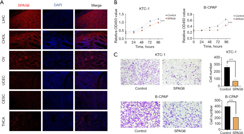
Figure 7.
Experimental validation of SPAG6 expression. (A) Multiple immunofluorescence staining of SPAG6 in pan-cancer tissue sections. Views of cancers, including high expression (LIHC, CHOL, and OV) and low expression (UCEC, CESC, and THCA) under microscopy (200×). SPAG6 = red, cellular nuclei = blue (DAPI). (B) Cell Counting Kit 8 assay of SPAG6. (C) Transwell migration assay of SPAG6 (10× magnification). The cells were fixed with 4% paraformaldehyde and stained with 0.1% crystal violet. Migrated cells were photographed with a microscope. ***, P<0.001. LIHC, liver hepatocellular carcinoma; CHOL, cholangiocarcinoma; OV, ovarian serous cystadenocarcinoma; UCEC, uterine corpus endometrial carcinoma; CESC, cervical squamous cell carcinoma and endocervical adenocarcinoma; THCA, thyroid carcinoma; DAPI, 4,6-diamidino-2-phenyiindole 2 hci.

