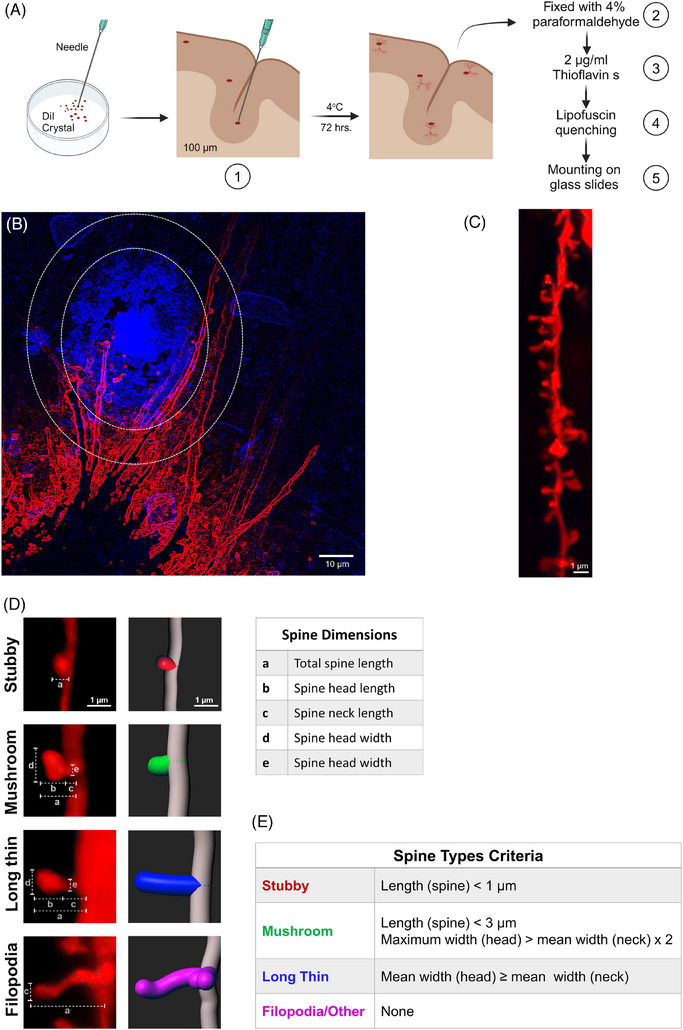
FIGURE 1.

Illustration of DiI labeling steps of neuronal structure in post mortem human frontal cortex and analysis of dendrites and spine parameters. (A) Simplified steps of DiI application on brain tissue sections and thioflavin labeling. (B) Representative picture of dendrites (red) surrounding the Aβ plaque (blue in a small oval). The area defined as proximal to the plaque includes Aβ plaque and the surrounding 10‐μm area. The area outside the large oval without plaque deposition is considered distal area. (C, D) Image of dendrite (C) and 3D reconstructions of the different types of spine (D) after analysis using Filament tracer and Classify Spines XTension Imaris analysis software version 9.9. Dendrite length, dendrite diameter, spine number, spine density, and dendritic spine morphology in the proximal area versus distal area were analyzed. (E) Spine classification criteria according to Classify Spines XTension Imaris analysis software version 9.9. Donor information described in Table 1A.
