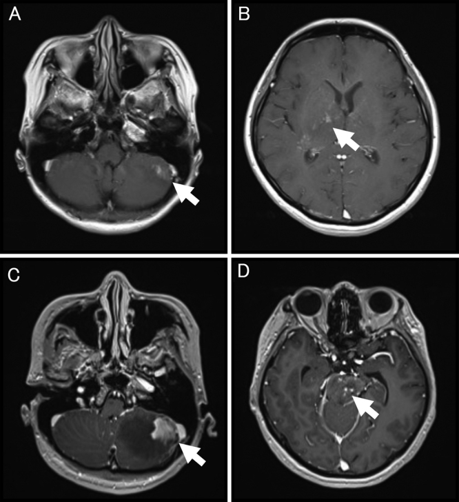
FIG. 1.

Axial gadolinium-enhanced T1-weighted MRI of the brain on admission (A and B) and 1 week before biopsy (C and D). MRI on admission shows abnormal enhancements scattered in the right basal ganglia (A, white arrow) and below the tentorium cerebelli (B, left cerebellum, white arrow). Images before biopsy show a lesion extending to the left cerebellum (C, white arrow) and new abnormal enhancement of the brainstem (D, white arrow).
