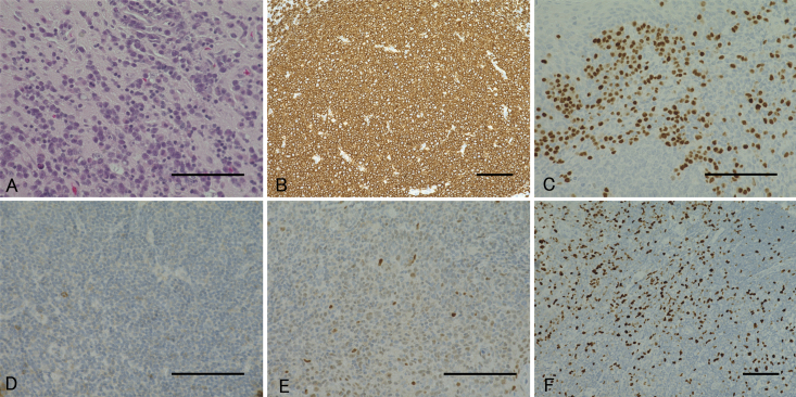
FIG. 2.
Pathological findings of the biopsied lesion. Hematoxylin and eosin staining shows marked diffuse infiltration of large lymphomatous cells (A), and immunostaining reveals these cells as CD20-positive (B), MUM-1–positive (C), CD10-negative (D), weakly positive for bcl-6 (E), and MIB-1–positive (F). Bar = 100 μm (A–F).

