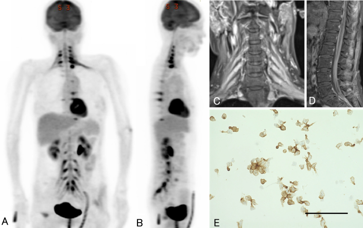
FIG. 3.
Images from FDG-PET, spinal MRI, and CSF cytology performed 25 months after initial diagnosis. Coronal (A) and sagittal (B) FDG-PET shows abnormal FDG accumulation in the ganglia, plexuses, and peripheral nerves from the cervical cord to the sacral cord. Coronal gadolinium-enhanced T1-weighted MRI of the cervical spinal cord (C) also shows multiple areas of contrast enhancement in the ganglia and plexuses. Sagittal gadolinium-enhanced T1-weighted MRI of the lumbar level (D) shows diffuse contrast effects within the spinal cavity. Immunostaining (E) shows aggregates of large, atypical lymphocytes positive for CD20 antibody. Bar = 100 μm.

