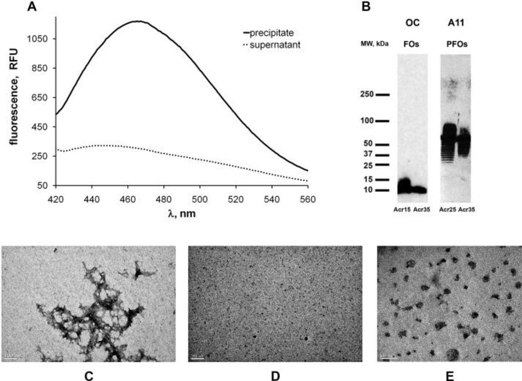
Fig. 1. Aβ40 aggregates and incorporation of acrylodan-labeled peptide.
(A) Acrylodan-labeled Aβ40 incorporates into amyloid fibrils. Amyloid fibrils prepared from 90% unlabeled Aβ40 and 10% Aβ40-Acr20 were collected by centrifugation (18,000 rpm, 1 h). Fluorescence spectroscopy of acrylodan indicated that Aβ40-Acr20 was present primarily in the fibril fraction. (B) Incorporation of 10% of acrylodan-labeled Aβ40 does not disrupt the structure of Aβ40 oligomers as indicated by Western blots. FOs labeled at positions 15 and 35 were detected with OC antibody and PFOs labeled in positions 25 and 35 were detected with A11 antibody. (C–E) Morphology of Aβ40 fibrils (C), FOs (D) and PFOs (E) determined by electron microscopy.

