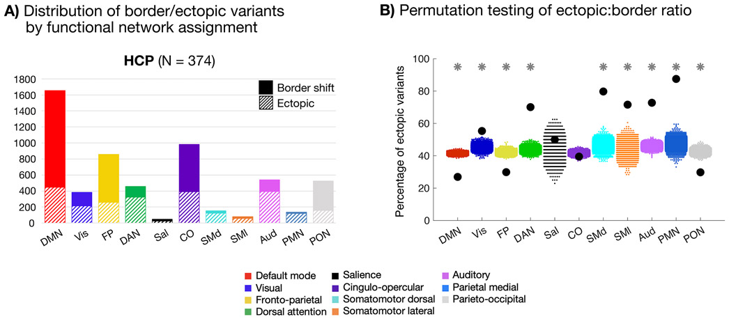
Fig. 4: Network linkages of border and ectopic variants.
(A) Network distributions of border and ectopic variants in the HCP dataset. Variants of both forms are commonly associated with the DMN, FP, and CO networks, as reported in past work15. Similar results were seen in the MSC dataset (Supp. Fig. 13) and using the parcellation-free approach to defining border and ectopic variants (Supp. Fig. 14) (B) Plot depicting permutation testing of the ectopic:border ratio in the HCP dataset. For all networks with the exception of salience and cingulo-opercular, the true proportion of ectopic variants (black dots) was significantly different from permuted proportions (colored dots, 1000 random permutations of shuffled labels) at p<0.001 (*; FDR corrected for multiple comparisons). DMN, FP, and PON variants were more likely to be border shifts, while sensorimotor, DAN, and PMN variants were more likely to be ectopic. Notably, ectopic variants were commonly found in all systems. See Supp. Fig. 12 for cortical depiction of each listed network.

