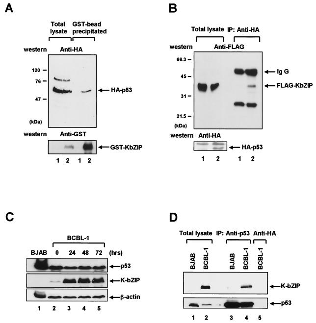
FIG. 2.
K-bZIP interacts with p53 in vivo. (A) 293T cells were transiently transfected with a plasmid encoding HA-p53 (pcDNA3/HA-p53) in combination with either GST (pEBG) (lanes labeled 1) or GST-KbZIP (pEBG/K-bZIP) (lanes labeled 2). Whole-cell lysates were prepared 48 h after transfection and were precipitated with glutathione-Sepharose beads. The bead-bound proteins were separated on a 7% polyacrylamide gel, transferred to a nitrocellulose membrane, and immunoblotted (Western) with anti-HA antibody (upper panel) or anti-GST antibody (lower panel). HA-p53 and GST-K-bZIP are indicated by arrows. (B) 293T cells were transfected with a plasmid encoding FLAG-KbZIP (pME18S/K-bZIP) in combination with either an HA-p53 expression plasmid (pcDNA3/HA-p53) (lanes labeled 2) or a control plasmid (pcDNA3) (lanes labeled 1). Cell lysates were subjected to immunoprecipitation (IP) with anti-HA monoclonal antibody. Immunoprecipitated proteins were separated on a 12% polyacrylamide gel, followed by immunoblotting (Western) with anti-FLAG antibody (upper panel). Immunoglobulin G (IgG) and Flag-K-bZIP are indicated by arrows. The identical membrane was reprobed with anti-HA antibody (lower panel) to confirm the expression of HA-p53 (indicated by an arrow). (C) The level of p53 and K-bZIP during the lytic replication cycle. BCBL-1 cells were treated with TPA as described previously (25). Whole cell lysates were prepared after the indicated number of hours, and Western blots were performed on equal quantities of whole cell extract (50 μg) by using p53 specific monoclonal antibody (upper panel), K-bZIP specific polyclonal antibody (middle panel), and β-actin antibody (lower panel). (D) The direct coimmunoprecipitation assay was performed using the BCBL-1 cell line 48 h after TPA induction. BCBL-1 cells and BJAB cells (107 cells) were harvested, and cell lysates were subjected to IP with p53 specific monoclonal antibody (lanes 3 and 4) and anti-HA antibody (lane 5). K-bZIP was detected using K-bZIP polyclonal antibody (upper panel), and the same membrane was reprobed with p53 antibody (bottom panel).

