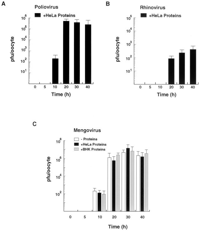
FIG. 3.
Time course studies of picornavirus replication in oocytes. (A) Poliovirus replication in Xenopus oocytes. Ten nanograms of poliovirus RNA was injected into oocytes together with 100 ng of HeLa cell cytoplasmic proteins and incubated at 30°C for 0, 5, 10, 20, 30, and 40 h. The titer of infectious poliovirus particles was determined by plaque assays in HeLa cells. (B) HRV 14 replication in Xenopus oocytes. The experiment was carried out under the conditions used for panel A. (C) Mengovirus replication in Xenopus oocytes. Ten nanograms of mengovirus RNA was microinjected into oocytes alone or together with 100 ng of cytoplasmic proteins from HeLa cells or BHK cells and incubated at 30°C for 0, 5, 10, 20, 30, and 40 h. Infectious-mengovirus titers were determined by plaque assays in BHK cells as for Fig. 2. The standard errors were calculated from three independent microinjections using the same batch of oocytes.

