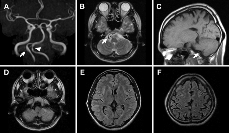
FIG. 1.
MRI was performed at 18:30 hours, after the patient had visited a private clinic on the 2nd day after the onset of headache. A: Magnetic resonance angiography (MRA) image showing a normal right VA (arrow) and mild stenosis (arrowhead) of the left VA. B: An axial T2-weighted image at the level of the VA shows a normal flow void sign. C: A sagittal T1-weighted image showing a flow void sign in the right VA without a T1-hyperintensity sign, indicating an intramural hematoma. D–F: Serial axial fluid-attenuated inversion recovery images showing no SAH at either the supra- or infratentorial cisterns.

