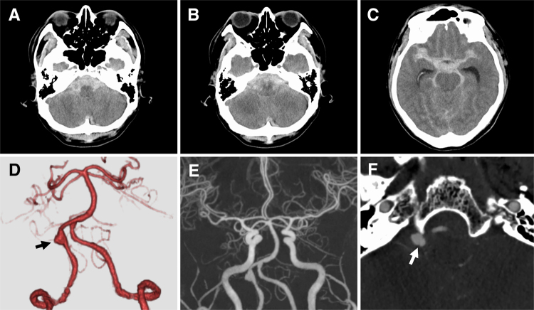
FIG. 2.
Imaging was performed at 23:00 hours, after the patient had been transported to our hospital via an ambulance. A–C: Emergency CT scans showing Fisher grade 3 SAH associated with acute hydrocephalus. A clot of SAH is predominant in the posterior fossa. D and E: CTA images showing aneurysmal dilatation of the right VA (arrow) and no other lesion suspected of dissection. F: The source image of the CTA image reveals an intimal flap (arrow).

