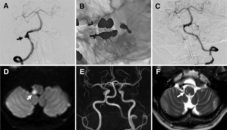
FIG. 3.
A: A digital subtraction angiography image showing aneurysmal dilatation (arrow) associated with a slight string sign at the proximal portion of the right VA. B: A nonsubtracted left oblique image obtained at the end of embolization, showing the coils that were placed during aneurysmal dilatation and in the right VA. C: A final left VA angiogram showing no opacification of the right VA or VADA. D: An axial diffusion-weighted image obtained on the day after the procedure, showing a right lateral medullary infarction (arrow). E: Two-year follow-up MRA image showing that there was no recurrence of the aneurysm and that the left VA had no abnormal findings. F: Axial T2-weighted image showing an old lateral medullary infarction (arrow).

