
FIGURE 1.
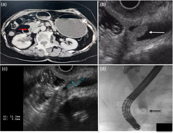
(a) Contrast‐enhanced abdominal computed tomography scan showing distal common bile duct polypoid lesion (red arrow). (b) Endoscopic ultrasound revealed a papillary mass with intraductal extension (white arrow). (c) Intraductal extension of the papillary mass measured 1.13 × 0.4 cm on endoscopic ultrasound. (d) Cholangiogram showed a filling defect within the papilla of Vater, extending proximally into the intraductal portion of the common bile duct.