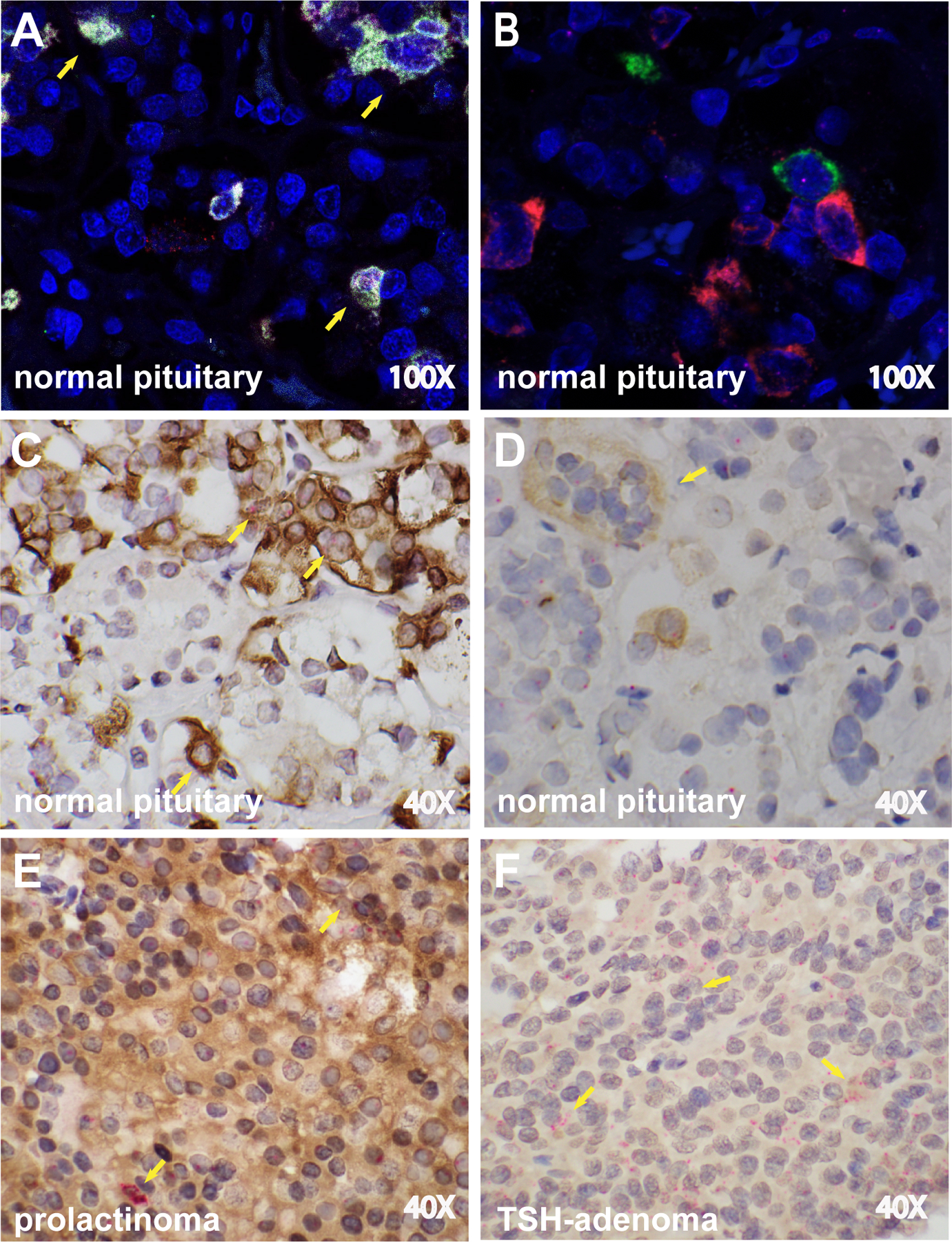
Figure 4. CTLA4 mRNA and protein is expressed in PRL-secreting cells in normal pituitary and prolactinoma.

(A) RNAscope Multiplex Fluorescent assay on a normal pituitary gland shows the simultaneous expression of PRL mRNA (white dots) and CTLA4 mRNA (red dots), as indicated by the yellow arrows, while (B) no colocalization is seen for TSH and CTLA4 mRNA. (C) Dual ISH/IHC for CTLA4 mRNA (red dots) and PRL protein (brown staining) in a normal pituitary. The co-expression of CTLA4 mRNA and PRL protein can be seen in multiple cells (yellow arrows). (D) Dual ISH/IHC for CTLA4 mRNA (red dots) and TSH protein (brown staining) in a normal pituitary. A cluster of TSH-secreting cells expressing CTLA4 is shown (yellow arrow). CTLA4 mRNA is also visible in other cells that do not secrete TSH. (E) Dual ISH/IHC for CTLA4 mRNA and PRL protein in a PRL-secreting tumor. Diffuse brown staining for PRL and multiple clusters of CTLA4 mRNA copies (yellow arrows). (F) Dual ISH/IHC for CTLA4 mRNA and TSH protein in a TSH-secreting tumor. Diffuse brown staining for TSH and multiple clusters of CTLA4 mRNA copies (yellow arrows). Original magnification times 100 (A and B) or 40 (C-F).
