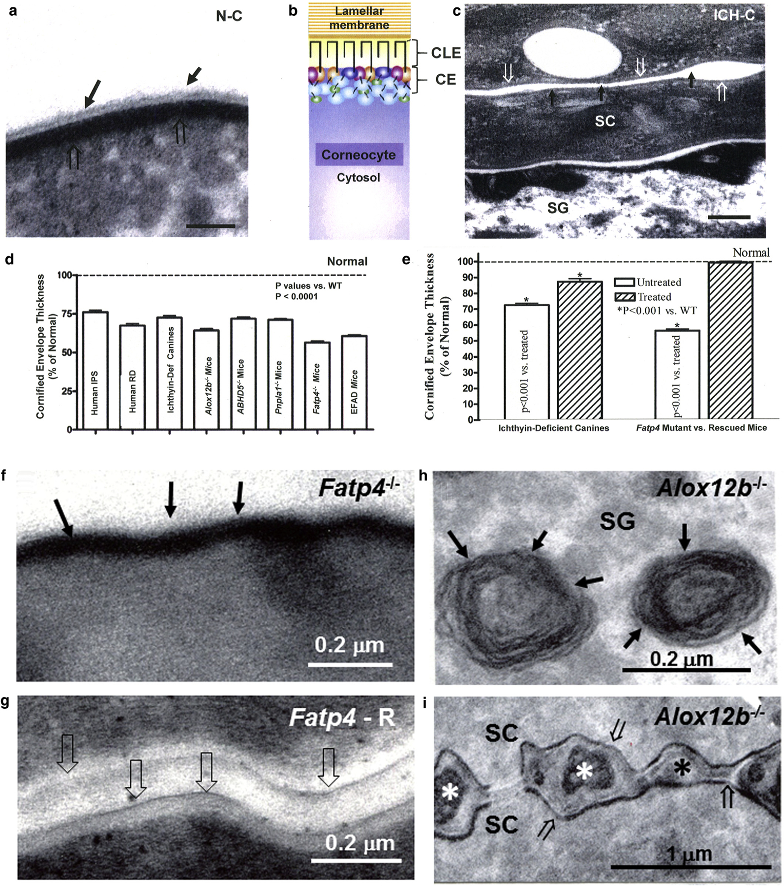
Figure 1. Dual scaffold functions of CLEs in several ARCIs.

(a, c) Loss of both CLE (solid arrows) and attenuation of cornified envelopes (CE) (open arrows in ichthyin-deficient canines [ICH-C] vs. replete CLEs and CEs in normal littermates [N-C]). (b) Model of the relationship between CLE, CE, lamellar membranes, and corneocyte cytosol in normal stratum corneum. (d, e) Significant decline in CE thickness in several ARCI patients and animal models (mean ± standard error of the mean). (e) Normalization of CE in ichthyin-deficient canines treated with topical ω-O-acylceramide versus vehicle alone. (f) Loss of CLE in Fatp4 knockout (−/−) (solid arrows) and (g) reappearance of CLE (open arrows) after suprabasal transgenic rescue (Fatp4-R) (see Mauldin et al. [2018] for images of CLE in ichthyin-deficient canines]. (h) Despite replete lamellar body contents (solid arrows), (i) secreted contents fail to transform into lamellar membranes in Alox12b−/− mice (asterisk). Note also the loss of CLEs and attenuation of CEs in Alox12b−/− SC (i, open arrows). (c, g, i) osmium tetroxide postfixation. (a, f, m) Ruthenium tetroxide postfixation after pyridine pretreatment. Scale bars = 100 nm in a; 0.2 µm in c, f, g, and h; and 1 µm in i. ARCI, autosomal recessive congenital ichthyosis; CE, cornified envelope; CLE, corneocyte lipid envelope; Def, deficient; IPS, ichthyosis prematurity syndrome; RD, Refsum disease; SC, stratum corneum; SG, stratum granulosum; WT, wild type.
