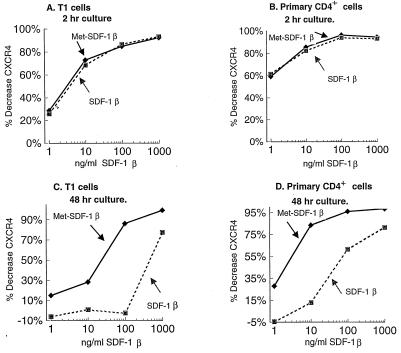
FIG. 6.
CXCR4 expression following culture with Met-SDF-1β versus wild-type SDF-1β. T1 cells (A and C) and primary CD4+ lymphocytes (B and D) were incubated with the indicated concentrations of Met-SDF-1β (⧫), SDF-1β (■), or medium without added chemokine. The expression level of CXCR4 was determined with anti-CXCR4 MAb (12G5) and flow cytometry following 2 h (A and B) or 48 h (C and D) of culture. The percent decrease of CXCR4 was calculated as (ΔMFI for cells cultured with chemokine)/(ΔMFI for control cells) × 100, as described in Materials and Methods. Similar results were obtained in multiple experiments (n > 3).

