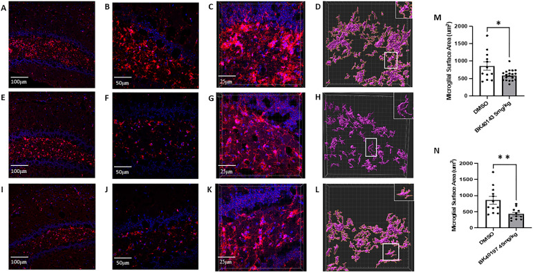
Figure 7. BK40143 and BK40197 alleviate microglial inflammatory morphology.
(A, B, C, E, F, G, I, J, K) Immunohistochemical staining of IBA1+ microglia revealed increased staining intensity in TgAPP mice treated with DMSO ((A) 20x, (B) 40x, (C) 63x z-stack) compared with mice treated with (E, F, G) 5 mg/kg BK40143 and (I, J, K) 45 mg/kg BK40197. (D, H, L, M, N) IMARIS 3D reconstruction and analysis revealed microglia from (D) DMSO-treated mice displayed significantly greater surface area than microglia from mice treated with (H, M) 5 mg/kg BK40143 (P = 0.01) and (L, N) 45 mg/kg BK40197 (P = 0.004).

