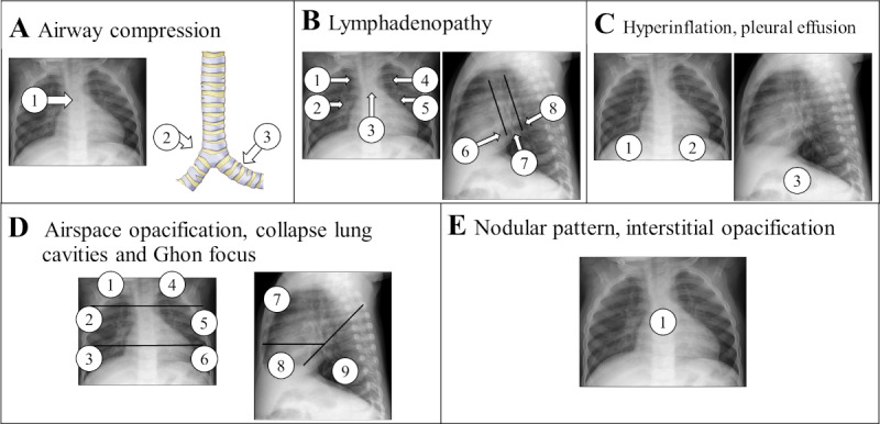
Figure 3.

Evaluation templates with the location of the specific findings that should be assessed by the evaluators with “yes” or “no” for each of the 10 sections. (A) Locations for the evaluation of possible airway compression or tracheal displacement. (B) Locations for the assessment of soft tissue density suggestive of lymphadenopathy. (C) Locations for the assessment of hyperinflation and pleural effusion. (D) Locations for the evaluation of air space opacification, collapsed lung, cavities, and calcified parenchyma. (E) Location for the assessment of nodular pattern, either miliary or larger widespread and bilateral nodules, and interstitial opacification. Based on [15,16] and chest x-ray review tool developed by Andronikou and the South African Tuberculosis Vaccine Initiative and used in Graham et al [16].
