
Figure 8.
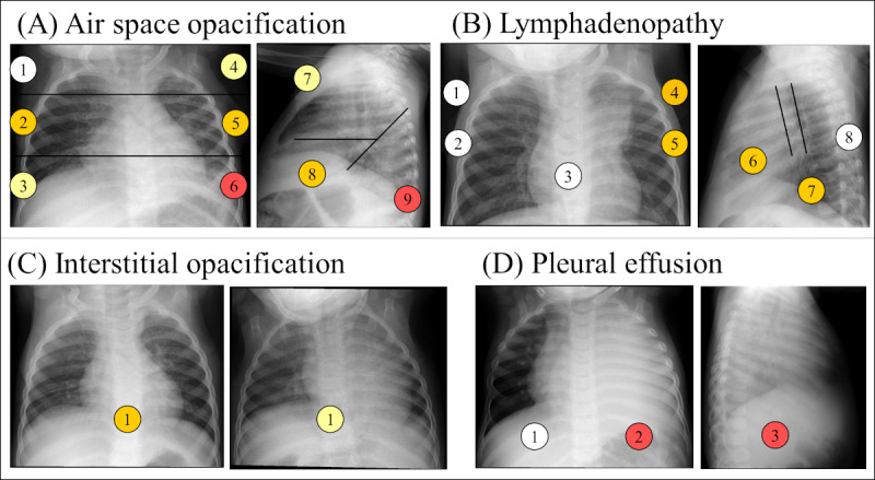
Example of evaluations of findings in different studies. The locations of the findings are defined in Figure 3. The color of the locations represents the number of evaluators that identified the presence of the finding in that location, being 0 evaluators for white, 1 evaluator for yellow, 2 evaluators for orange, and 3 evaluators for red. (A) Presence of air space opacification in the anteroposterior (AP) and lateral chest x-ray (CXR) views of an examination of a female patient of 11 months classified as unconfirmed tuberculosis (TB) and as suggestive of TB by 1 out of the 3 evaluators. (B) Presence of lymphadenopathy in the AP and lateral CXR views of an examination of a female patient of 11 months classified as confirmed TB and as suggestive of TB by the 3 evaluators. (C) Presence of interstitial opacification on AP CXR views of 2 studies, the one on the left is from a male patient of 1 year and 4 months of age. Both studies were classified as unconfirmed TB and not suggestive of TB. The AP view on the right corresponds to a patient of female of 11 months of age. The examination was classified as unlikely TB and 1 out of 3 evaluators assessed it as confirmed TB. (D) Presence of pleural effusion in the AP and lateral view of an examination of a male of 2 years and 2 months of age classified as confirmed TB and evaluated as suggestive of TB by the 3 evaluators.