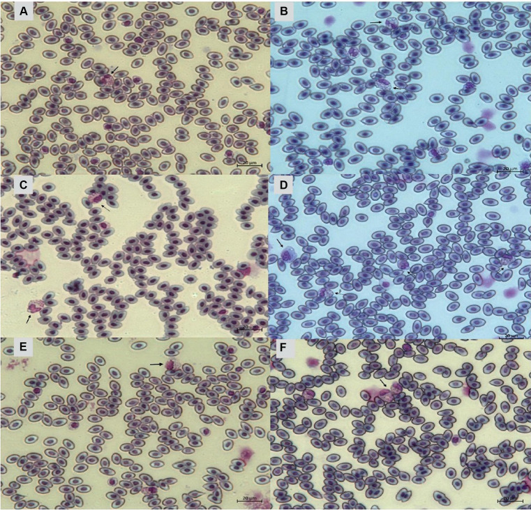
Figure 11.
Heterophils of birds (Gallus gallus domesticus) in four poultry houses (PH) in the Southwest of Mato Grosso do Sul, Brazil. Cell morphology were evaluated according to Table 3 for comparative purposes. Images (A,B) represents heterophil of the classification 3, severe alteration (reversible lesion). Images (C–F) represents heterophils abnormal of the classification 4, severe alteration (irreversible lesion). Methanolic Wright-Giemsa stain. The hematological slides were analyzed using an Optical Microscope (MOC), Carl Zeiss Microscopy GmbH, Axio Scope Al model, with the assistance of ZEN Lite (Blue Edition) imaging software, at magnifications of 1,000×. The images of the smears were photographed using an Axiocam 503 color camera connected to the MOC.

