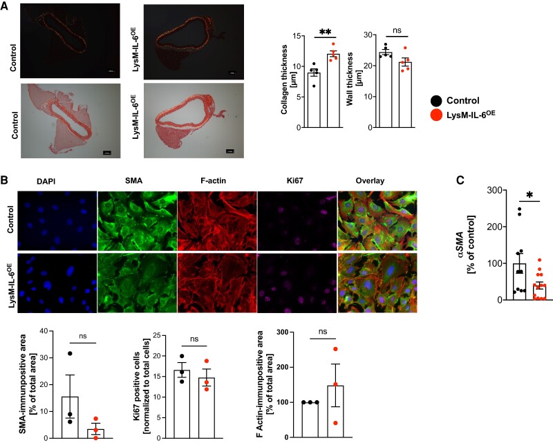
Figure 4.
Interleukin-6-induced vascular dysfunction is associated with an altered vascular smooth muscle cell phenotype and fibrosis formation. (A) Collagen deposition in aortic sections. Sirius Red staining of aortic sections. Left: representative image of aortic sections (images without polarized light are shown below), scale bar = 50 µm. Right: aortic wall thickness and collagen thickness measurement at 10 different points/section with ImageJ software, n = 5, unpaired Student’s t-test. P = 0.0079 (collagen thickness). (B) Immunocytochemical analysis of vascular smooth muscle cell phenotype. Vascular smooth muscle cells were isolated from LysM-IL-6OE mice and control mice and cultured. Cultured vascular smooth muscle cell were stained with smooth muscle alpha-actin, F-actin, Ki67, and DAPI (60×). Representative images are shown in the top row. Quantification is shown in the bottom row. n = 3 mice per group of two independent experiments. Single images of three biological replicates per mouse were analysed, and the mean values each mouse were compared, unpaired Student’s t-test. (C) Quantitative real-time PCR analysis for alpha SMA in LysM-IL-6OE mice aortas (red) vs. control aortas. Housekeeping gene: Gapdh. n = 10–12 mice per group, unpaired Student’s t-test. P = 0.03. Data are presented as mean ± SEM, and P values of <0.05 were considered significant and marked by asterisks (*P < 0.05; **P < 0.01; ***P < 0.001).

