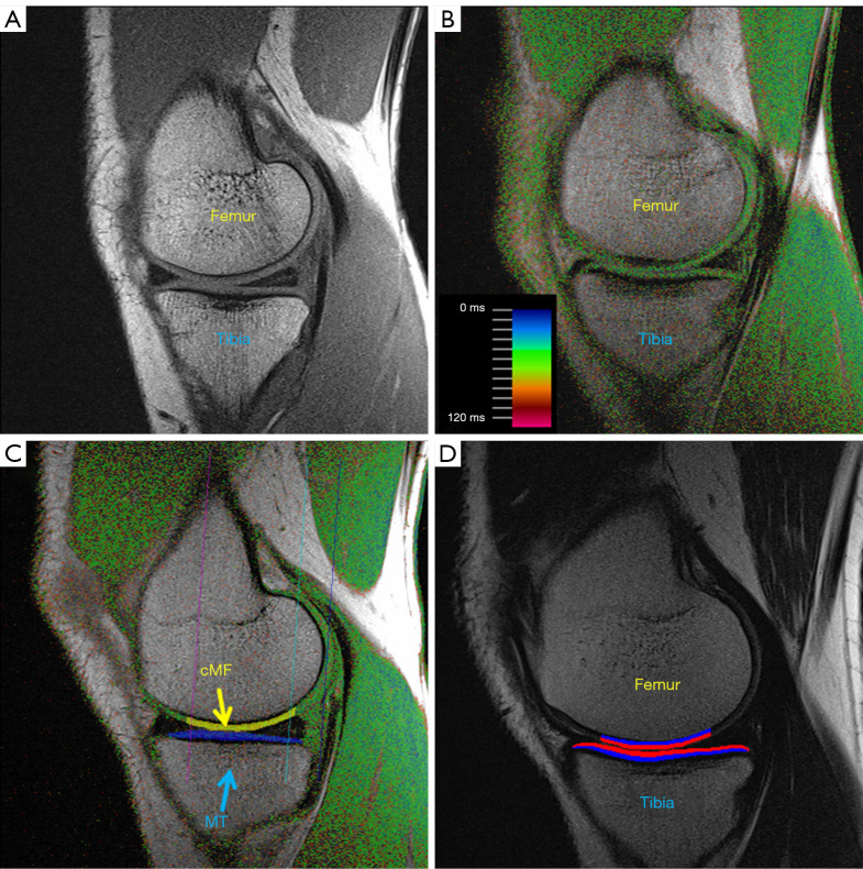
Figure 1.
MESE MR images of the MFTC in various study participant groups, with and without cartilage segmentation: (A) first echo of the MESE; Healthy Control Subject; (B) T2 map derived from the 7 echoes of the MESE (color coding provided); patient without knee instability: coper; (C) MESE with fully automated CNN-segmentation of the MT and cMF cartilage; patient with dynamic knee instability: non-coper; (D) MESE with fully automated CNN-segmentation, displaying the superficial 50% and deep 50% of the femorotibial cartilage plates; patient with surgical anterior cruciate ligament reconstruction. cMF, central (weight-bearing) medial femur; MT, medial tibia; MESE, multi echo spin echo; MR, magnetic resonance; MFTC, medial femorotibial compartment; T2, transverse relaxation time; CNN, convolutional neural network.

