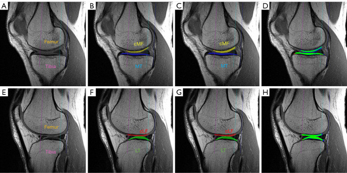
Figure 2.
Visual comparison of fully automated (7-echoes CNN) vs. manual segmentation in the medial (A-D) and lateral (E-H) femorotibial compartment. (A) Sagittal MESE MRI showing the (medial) tibial and femoral bone without cartilage segmentation; (B) manual segmentation in the medial compartment (tibia and femur); (C) automated segmentation in the medial compartment; (D) difference between manual vs. automated segmentation in the medial compartment; (E) sagittal MESE MRI showing the (lateral) tibial and femoral bone without cartilage segmentation; (F) manual segmentation in the lateral compartment (tibia and femur); (G) automated segmentation in the lateral compartment; (H) difference between manual and automated segmentation in the lateral compartment. (D) and (H) show both local underestimation (red color) and overestimation (blue) of the automated segmentations. Green color indicates agreement of both segmentation methods. cMF, central (weight-bearing) medial femur; MT, medial tibia; cLF, central (weight-bearing) lateral femur; LT, lateral tibia; CNN, convolutional neural network; MESE, multi echo spin echo; MRI, magnetic resonance imaging.

