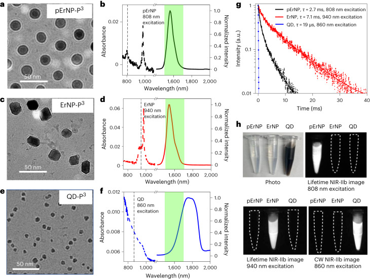
Fig. 1. Three nanoparticle probes luminescence/emitting in the 1,500–1,700 nm range for vaccine carrier and multiplexed NIR/SWIR imaging.
a,c,e, Cryo-EM images of pErNP-P3 (a), ErNP-P3 (b) and QD-P3 (e) in PBS buffer. These images show a uniform coating of approximately 5 nm thickness on the three types of particle. b,d,f, Absorption (dashed curves) and emission (solid curves) spectra of three different nanoparticles, pErNP-P3 (b), ErNP-P3 (d) and QD-P3 (f). The green shaded areas are the NIR-IIb 1,500–1,700 nm detection range for imaging. The dashed vertical lines were drawn to show the excitation wavelengths (808 nm for pErNP, 940 nm for ErNP and 860 nm for QD) used for three-plex imaging for each of the three probes in lifetime (for pErNP and ErNP) or CW (for QD) mode. g, Lifetime decays of pErNP, ErNP and QD, respectively. In b, d and f, pErNP and ErNP were measured in cyclohexane, QD was measured in PBS buffer, and photoluminescence (PL) decay curves were fit with a typical bi-exponential function. h, A colour photograph and multiplexed NIR-IIb imaging of pErNP, ErNP and QD in PBS buffer under three different imaging conditions that detect one probe at a time without imaging the other two. pErNP: 808 nm excitation, 1,500–1,700 nm detection in lifetime mode (for three-plex imaging details, see Supplementary Information). ErNP: 940 nm excitation, 1,500–1,700 nm detection in lifetime mode; QD: 860 nm excitation, 1,500–1,700 nm in CW.

