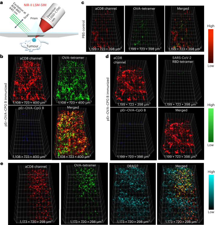
Fig. 5. In vivo 3D volumetric NIR-II LSM-SIM imaging of antigen specific CD8+ CTLs in tumour microenvironment.
a, Scheme of in vivo NIR-II LSM-SIM imaging18 with illumination and detection at 45° to the tumour (for details, see Supplementary Information). b, Three-dimensional volumetric NIR-II SIM images of ErNP–aCD8 (red), and QD–OVA–tetramer (green) recorded in the tumour in the pErNP–OVA–CpG B nanovaccine immunized mouse 48 h after i.v. injection of ErNP–aCD8, and QD–OVA–tetramer. It is important to note that the pEr signal is weak at this timepoint. c, The same as in b except that the mouse was ‘immunized’ by PBS buffer. d, The same as in b (mouse immunized with pErNP–OVA–CpG B nanovaccine) except that QD–RBD–tetramer was used instead of the QD–OVA–tetramer. e, Three-dimensional volumetric NIR-II SIM images of tumour tissue ex vivo. After in vivo SIM imaging, the mouse was killed under anaesthesia and the tumour was removed. The tumour was fixed in 10% neutral-buffered formalin for 30 min at room temperature, then washed three times with 1× PBS buffer and labelled with the nuclear dye, DRAQ7, for 3 h. After washing the tumour three times with 1× PBS buffer, it was preserved in glycerol at 4 °C for ex vivo imaging. Imaging conditions for ErNP: 940 nm excitation, 1,500–1,700 nm detection, exposure times 20 ms, lifetime mode; QD: 860 nm excitation, 1,500–1,700 nm detection, exposure times 100 ms, CW mode; pErNP: 808 nm excitation, 1,500–1,700 nm detection, exposure times 20 ms, lifetime mode; DRAQ7: 650 nm excitation, 690–850 nm detection, exposure times 100 ms, CW mode.

