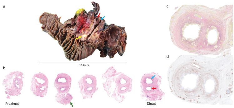
Fig. 2.
(a) Macroscopic photograph of the pancreaticoduodenectomy specimen, demonstrates a duplicated common bile duct as indicated by the red (right duct) and blue (left duct) arrows. The yellow arrow points to the ampullary tumor. (b) Serial sections from the duplicated common bile duct. The green arrow highlights the cystic duct adjacent to the right-sided duplicated bile duct. The bile duct at the proximal resection margin was single (HE, x4). (c) Elastin van Gieson stain and (d) immunohistochemistry for smooth muscle actin showing complete layering by smooth muscle of each individual duct (x10).

