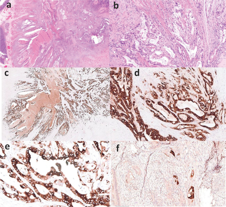
Fig. 3.
Histology and immunohistochemistry of ampullary adenocarcinoma (a – HE, x14) and (b – HE, x20) with positive expression for CK7 (c – IHC, anti-CK7 mAb, x20), epithelial membrane antigen (d – IHC, anti-EMA mAb, x20) and MUC5AC (E – IHC, anti-MUC5AC, x400). There is focal squamous differentiation highlighted by CK5/6 (F – IHC, anti-CK5/6, x20).

