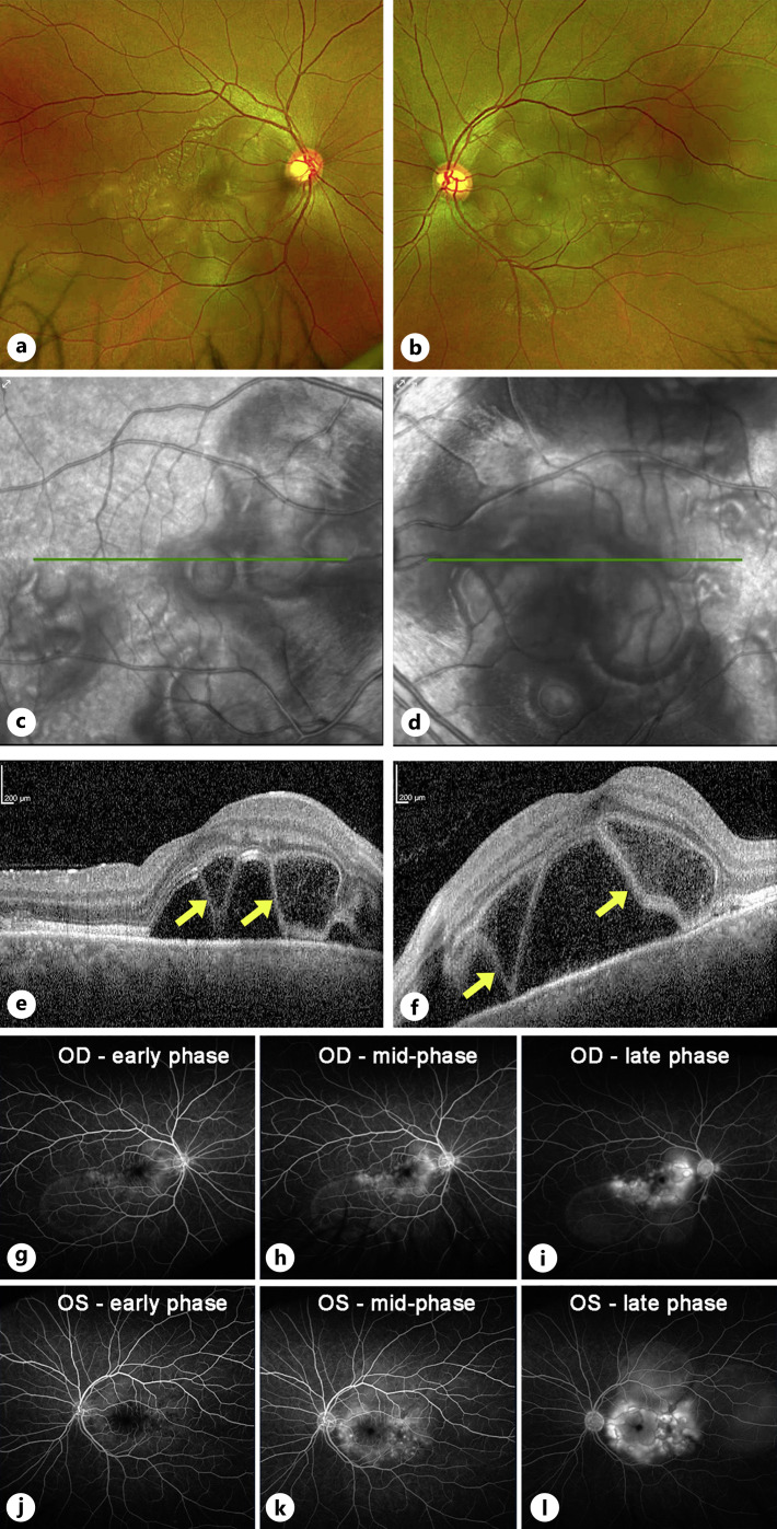
Fig. 1.
Imaging at presentation. Color fundus photos of the right (a) and left (b) eyes demonstrate bilateral posterior pole multiloculated subretinal fluid pockets. There is no evidence of disc edema, hyperemia, or any vascular sheathing, and the media is clear. Infrared images (c, d) better highlight the bilateral multiloculated pockets of subretinal fluid. Optical coherence tomography (e, f) of the macula showed a prominent accumulation of outer retinal and subretinal fluid, along with areas of bacillary layer detachments (BALAD, yellow arrows). Fluorescein angiography demonstrated scattered areas of leakage within the macula and multiloculated areas of pooling in both the right (g–i) and left (j–l) eyes. No leakage is noted from the optic discs or vessels.

