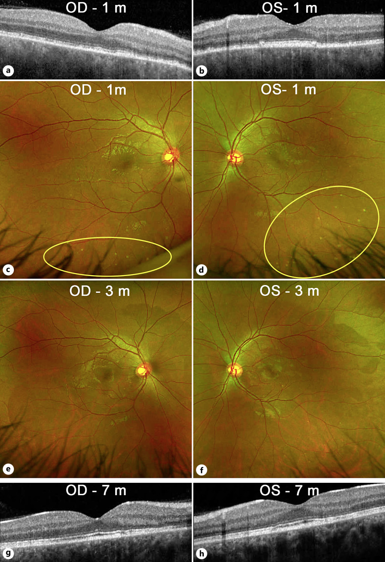
Fig. 4.
Imaging after the acute phase. a, b Optical coherence tomography of the macula at 1 month showed complete resolution of the macular fluid and bacillary layer detachments OU. Some residual ellipsoid zone, interdigitation zone, and RPE changes can be seen. c, d Color fundus photos showed resolution of the subretinal fluid blebs, but a new finding of white/yellow dots in the inferior midperiphery OU (yellow ovals). e, f By 3 months, these white dots had mostly resolved. g, h No recurrence of macular fluid was seen up to 7 months of follow-up on OCT. Some improvement of the ellipsoid changes was seen in both eyes.

