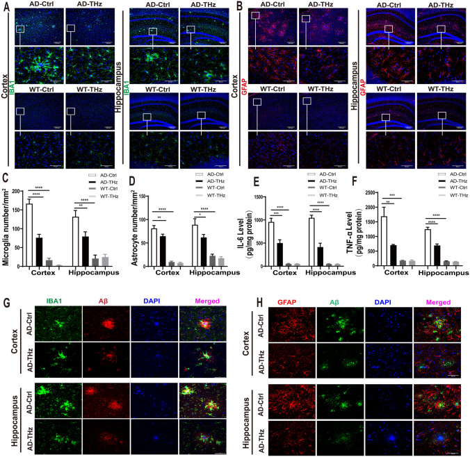
Fig. 6.
THz waves suppress neuroinflammation in the brain of APPSWE/PS1DE9 mice. THz waves decrease glial reactivity in APPSWE/PS1DE9 mice. A Representative immunostaining of IBA-1 (green) in the cortex and hippocampus (Hip) of the different groups. Scale bars, 200 µm, 50 µm. B Representative immunostaining of GFAP (red) in the cortex and hippocampus of the different groups. Insets: representative morphology at higher magnification. Scale bars, 200 µm. C Numbers of microglia in the cortex and hippocampus. D Numbers of astrocytes in the cortex and hippocampus. n = 6 mice per group for AD, n = 3 mice per group for WT. E, F THz waves decrease the of production pro-inflammatory cytokines IL-6 and TNF-α in the brain of APPSWE/PS1DE9 mice, measured by ELISA. n = 6 per group for AD, n = 4 mice per group for WT. G Colocalization of fluorescent Aβ (red), Iba1 (green), and DAPI (blue) in the cerebral cortex and hippocampus. Scale bar, 50 μm. H Colocalization of fluorescent Aβ (green), GFAP (red), and DAPI (blue) in the cerebral cortex and hippocampus. Scale bar, 50 µm. Data are presented as the mean ± SEM. ****P <0.0001, ***P <0.001, **P <0.01. AD APPSWE/PS1DE9, transgenic mice; Ctrl, control; WT. wild type.

