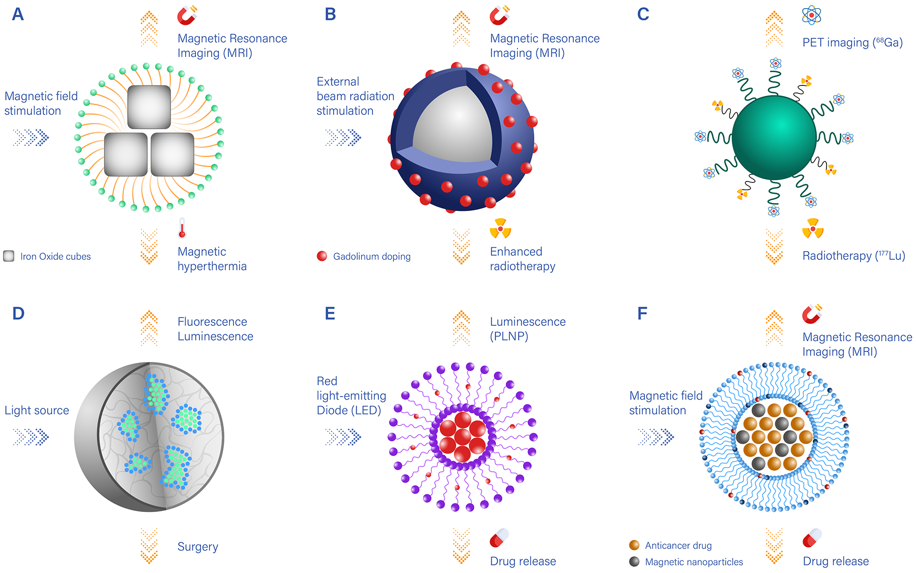
Figure 2 |. Examples of nanotheranostics with different imaging, therapeutic and targeting components.

A. Clusters of iron oxide nanocubes coated by polymeric or lipid chains (yellow) and presenting surface-targeting moieties (blue). Upon magnetic stimulation, iron oxide nanocubes generate thermal energy, which can be used for magnetic hyperthermia, and can be visualized via magnetic resonance imaging (MRI); B. Nanocarriers carrying heavy metals (such as Gd) locally enhance the therapeutic efficacy of external beam radiation and can be imaged via MRI; C. Nanocarriers conjugated to two different radionuclides can be used for positron emission tomography (PET) imaging (using 89Zr) and radiotherapy (using 177Lu); D. Nanoparticles encapsulating luminescent/fluorescent molecules, which under external light stimulation can be visualised and guide surgical resection; E. Nanocarriers loaded with persistent luminescence nanoparticles (PLNP) and chemotherapeutic drugs that are released by passive diffusion over time; F. Nanocarriers loaded with iron oxide particles for MR imaging guidance, which under high-intensity focused ultrasound stimulation trigger the release of anti-cancer molecules.
