Fig. 1.
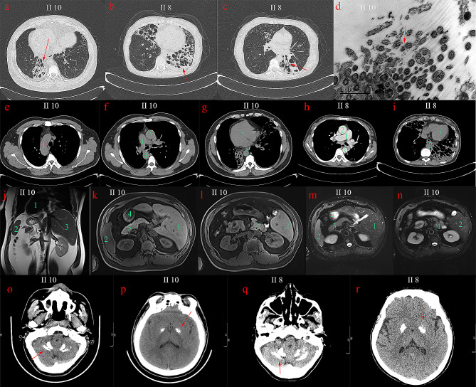
(a) Chest CT images of proband II10: the cystic and columnar expansion of the both lower lung bronchi. (b) Chest CT images of II8: the cystic dilatation of left lower lung and right middle lung bronchi. (c) bronchial cystic dilation and mucus filling in the left lower lung, tree-in-bud pattern was seen in the middle lobe of the right lung. (d) Bronchial mucosa of II10 was observed by electron microscopy, the ultrastructure of cilia was disordered, some microtubules were arranged in disorder, surrounding microtubules were reduced. (e) The right aortic arch of II10. (f) The right pulmonary aorta (1), right ascending aorta (2) and descending aorta (3) of II10. (g) The dextrocardia of II10. (h) The normal positions of pulmonary trunk, ascending aorta and descending aorta of II8. (i) The normal position of heart of II8; (j-n) Abdominal CT images of II10: the reversal of the position of the heart, liver, spleen, pancreas, and stomach. (o-r) Brain CT images of II10 and II8. o, q, Bilateral calcifications observed in dentate nucleus; p, r, bilateral and symmetrical calcifications observed in globus pallidus

