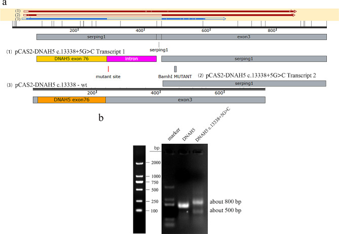
Fig. 3.
Minigene splicing assay The splicing pattern of DNAH5 WT was consistent with the report of NCBI, while DNAH5 mutation c.13,338 + 5G > C showed two types of splicing, with the target band of about 800 bp and 500 bp. The splicing form of 800 bp band was that some bases of the intron where the mutation site is located were translated; The splicing form of 500 bp band was the deletion of exon 76 where the c.13,338 is located. (a) A diagram of splicing: (1), (2) and (3) represent three forms of splicing respectively. (b) Agarose gel results of splicing modes of DNAH5 mutation c.13,338 + 5G > C and wild-type; full-length gels are presented in Supplementary Figure

