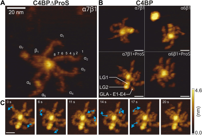
Figure 4. C4BP higher-order structures and their flexibility visualized by high-speed atomic force microscopy in a strong immobilization buffer.
(A) C4BP α7β1 (C4BP∆ProS sample). A central core with a diameter of ~5 nm surrounded by 7 C4BPα “arms” and a shorter C4BPβ chain. The C4BPα, C4BPβ, and 8 CCP domains of α2 were annotated. (B) Representative images of the four major species identified in human serum C4BP sample (α6β1 and α7β1 variants with or without ProS). (C) HS-AFM time series following a single C4BP molecule in a strong immobilization buffer highlighting the flexibility of C4BPα. Domains moving between frames are indicated by blue arrows. All scale bars correspond to 20 nm. Source data are available online for this figure.

