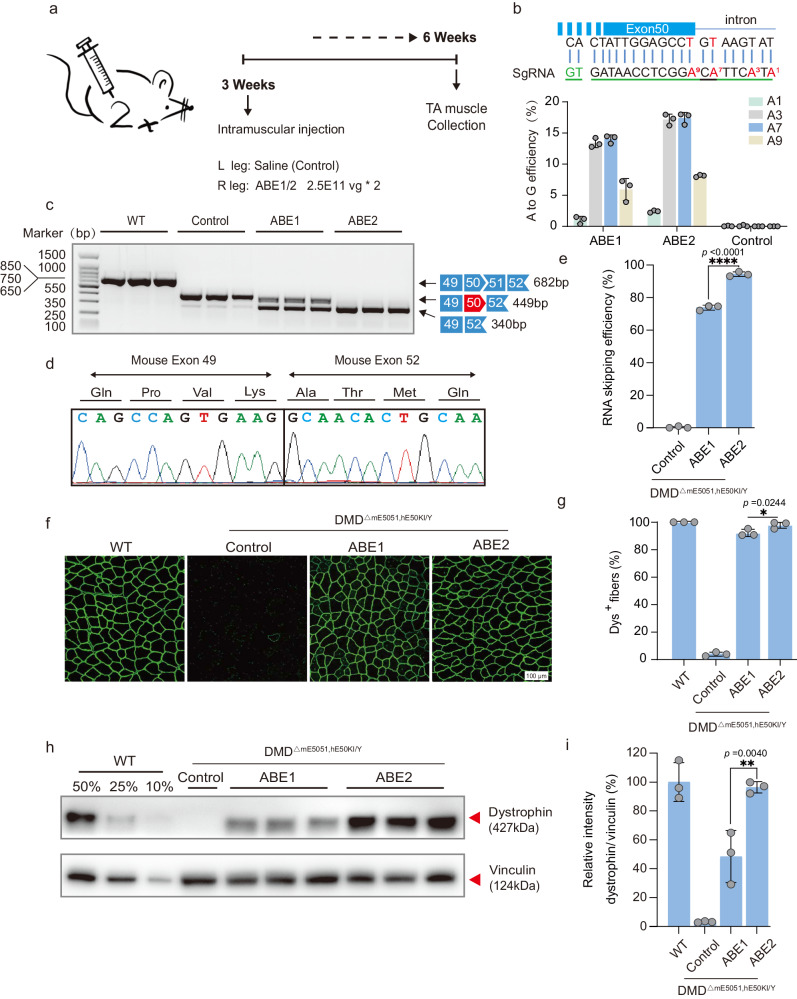
Fig. 3. ABE systems robustly rescued dystrophin expression in TA 6 weeks after local AAV injection.
a Overview for the in vivo intramuscular injection of the adeno-associated virus (AAV) -ABE construct into the tibialis anterior muscle of the right leg of 3-week-old DMDΔmE5051,KIhE50/Y mice. Left leg was injected with saline as a control. Black arrows indicate time points for tissue collection after injection. b The base editing efficiency of ABE1 and ABE2 were analyzed by deep sequencing. Sequence shows sgRNA (green line) and PAM (green letters). c RT-PCR products from muscle of DMDΔmE5051,KIhE50/Y mice were analyzed by gel electrophoresis. d RNA exon skipping was validated by Sanger sequencing. e Exon skipping efficiency by analyzing of total mRNA extracted from muscle tissue with deep sequencing. f Comparison of dystrophin (Dys+) expression restored by ABE1 and ABE2 systems with immunofluorescence staining. Dystrophin is shown in green. Scale bar, 100 μm. g Quantification of Dys+ fibers in cross sections of TA muscles. h Western blot analysis of dystrophin and vinculin expression in TA muscles 6 weeks after injection with ABEs or saline. i Quantification of dystrophin expression from Western blots after normalization to vinculin expression. Age-matched wild-type (WT) and saline-treated DMDΔmE5051,KIhE50/Y mice were included as positive and negative control Data are presented as mean ± s.d (n = 3 independent biological replicates). Each dot represents an individual mouse. Significance is indicated by asterisk and determined using unpaired two-tailed Student’s t test, *P < 0.05. **P < 0.01. ****P < 0.0001, NS represents not statistically significant. Source data are provided as a Source Data file.

