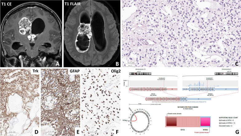
Fig. 1.
A, B Patient #2 (15 y/F) presented with an intraventricular diffuse low-grade glioma located at the lateral ventricle, exhibiting heterogeneous high signal intensity on contrast-enhanced T1-weighted and T1-fluid-attenuated inversion recovery (FLAIR) images. B The tissue in the H&E section resembled a myxoid glioneuronal tumour, characterized by monotonous round cells within a myxoid background. D, F Immunohistochemical analysis revealed positivity for GFAP, TRK, and Olig2 in this tumour. G An Arriba plot generated from next-generation sequencing (NGS) data using RNA identified the KIF5A::NTRK2 in-frame fusion in the tumour (C, D: H&E, E: GFAP, F: Olig2; scale bar, C-E: 200 μm, F: 100 μm)

