Fig. 2.
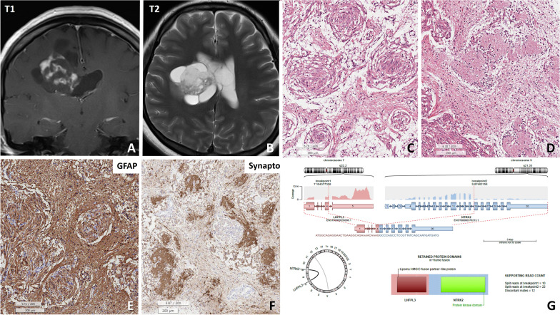
Patient #5, a 27-year-old female, presented with an uncommon glioma harbouring the LHFPL3::NTRK2 fusion. A, B T1 and T2 FLAIR MR images revealed a 5.3 × 4.5 × 1.6 cm solid and cystic mass in the right lateral ventricle with multifocal enhancement in the right intra- and periventricular white matter. C, D H&E sections of the tumour exhibited a distinctive pathology characterized by multiple whorls formed by glial cells and neuropil-like islands. E The spindle-shaped glial cells forming the whorls were positive for GFAP and negative for synaptophysin, but F the neuropil-like islands were positive for synaptophysin and negative for GFAP. G An Arriba plot generated from next-generation sequencing (NGS) data for tumour RNA identified the LHFPL3::NTRK2 in-frame fusion (scale bar, C–E: 200 μm, F: 100 μm)

