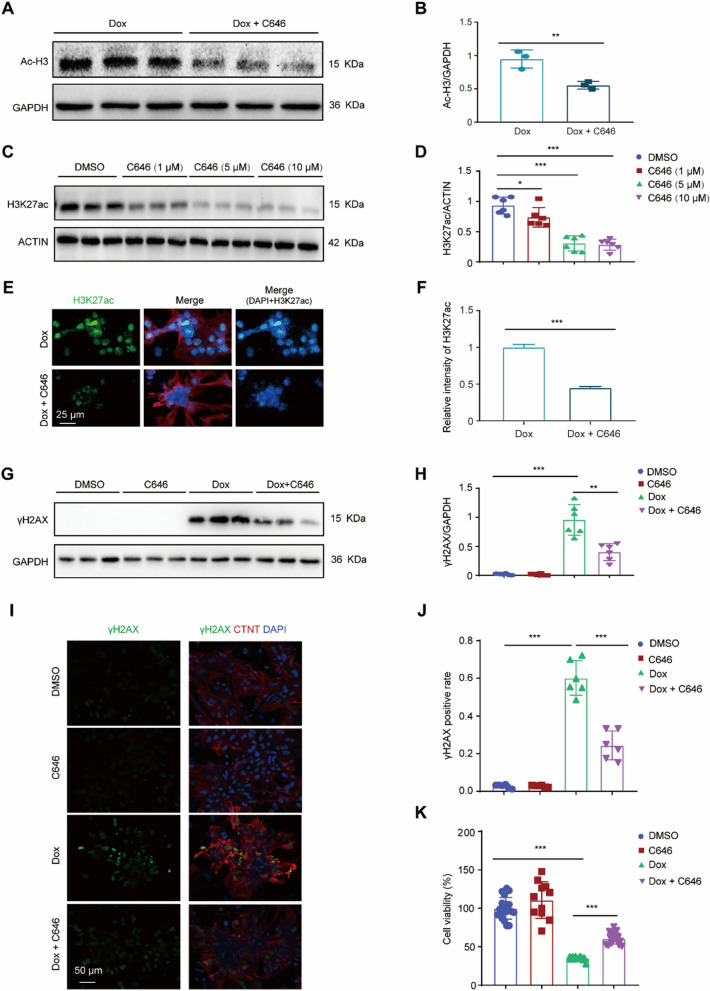
Fig. 2.
Reducing H3K27ac accumulation via C646 ameliorated Dox-induced DNA damage in cardiomyocytes. A, B Representative western blot (A) and statistical data (B) showing the levels of Ac-H3 from the cardiomyocytes treated with Dox or Dox with C646 for 24 h. n = 3 per group. C, D Representative western blot (C) and statistical data (D) showing the levels of H3K27ac from the cardiomyocytes treated with increasing concentrations of C646 ranging from 0 to 10 μM for 24 h. n = 6 per group. E, F Representative immunostaining image (E) and relative fluorescence intensity (F) for H3K27ac (green) in the cardiomyocytes treated with Dox or Dox with C646 for 24 h; red field denotes cardiac troponin T; Nuclei were stained with DAPI (blue). G, H Representative western blot (G) and statistical data (H) showing the levels of γH2AX from Dox-treated cardiomyocytes with or without C646 for 24 h. n = 6 per group. I, J Representative immunostaining image and analysis for γH2AX (green) in Dox-treated cardiomyocytes with or without C646 for 24 h. K CCK-8 assay revealed cell viability after Dox treatment with or without C646, respectively

