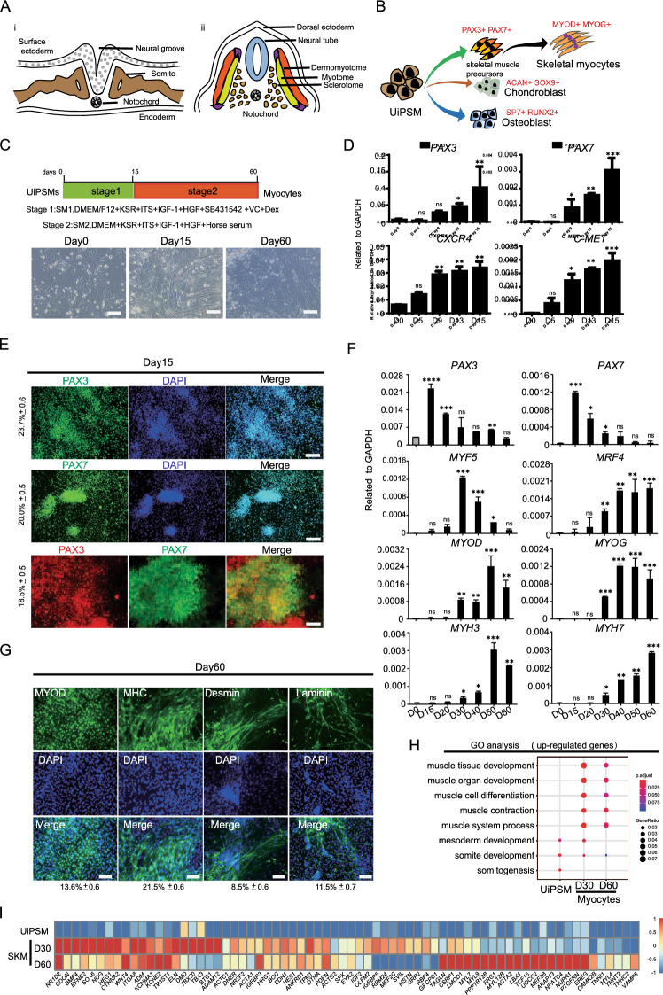
Fig. 1.
Differentiation of UiPSM cells into skeletal myocytes in vitro. A Schematic diagram of somite development. a. Illustration of the epithelial somite's spatial relationship to surrounding structure. b. Depiction of the differentiated somite's spatial relationship to surrounding structures. Dorsally, the somite differentiates into the dermomyotome and sclerotome. The dermomyotome subsequently gives rise to the myotome, which develops into skeletal muscle tissue. The sclerotome differentiates into osteoblasts and chondroblasts, forming the axial skeleton. B Schematic diagram of UiPSM cell differentiation into skeletal myocytes, osteoblasts and chondroblasts. C Schematic overview of stepwise differentiation of skeletal myocytes from UiPSM cells. Representative images show the morphological changes from UiPSM cells to skeletal muscle filaments. Scale bars, 100 µm. D Representative gene expression of human skeletal muscle satellite cells (PAX3, PAX7, CXCR4, C-MET) at day 15. Data are mean ± SD, n = 3 independent experiments. (*P ≤ 0.05). E Immunofluorescence of PAX3 and PAX7 during the differentiation of skeletal muscle cell form UiPSM at day 60 (left). The scale bar represents 100 µm. The values on the left represent the percentage of positive cells statistically. Data are mean ± SD, n = 3 independent experiments, each experiment counted 100 fields of view. F Representative gene expression of human skeletal muscle satellite cells (PAX3, PAX7) and skeletal myoblasts (MYOD, MYOG, MRF4) and skeletal myocytes (MYH3, MYH7) during the differentiation process. Data are mean ± SD, n = 3 independent experiments. (*P ≤ 0.05). G Immunofluorescence of MYOD, MHC, Desmin, Laminin during the differentiation of skeletal muscle cell form UiPSM at day 60 (left). The scale bar represents 100 µm. The following values represent the percentage of positive cells statistically. Data are mean ± SD, n = 3 independent experiments, each experiment counted 100 fields of view. H The UiPSM cells differentiated at day 30 and day 60 were enriched for GO terms of skeletal muscle development. I Heatmap illustrating the gene expression of skeletal muscle development related genes with dramatical change in UiPSM cell derived myocytes at day 30 and day 60

