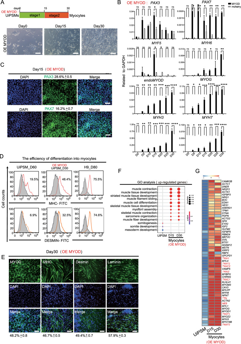
Fig. 2.
MYOD promoted the maturity of skeletal myocytes in vitro. A Schematic overview of stepwise differentiation of skeletal myocytes from UiPSM with ectopic MYOD. SKM: skeletal muscle cells. Representative images show the morphological changes from UiPSM cells to skeletal myocytes. Scale bars, 100 µm. n = 3 independent experiments. B Representative gene expression of human skeletal muscle satellite cells (PAX3, PAX7), skeletal myoblasts (MYOD, MYOG, MRF4) and skeletal myocytes (MYH3, MYH7) when overexpressed ectopic MYOD during the differentiation process. Mcherry as a negative control of overexpression vector. Data are mean ± SD, n = 3 independent experiments. (*P ≤ 0.05). C Detection of skeletal muscle satellite cell-specific genes (PAX3 and PAX7) in MYOD-mediated differentiated UiPSM cells at day 15. The following values indicate the percentage of positive cells statistically (Data are mean ± SD, n = 3 independent experiments). Scale bars, 100 µm. D Flow cytometric analysis evaluating differentiation efficiency via MHC and Desmin protein expression in skeletal muscle cells at day 60 of differentiation. hESC (H9)-derived skeletal muscle cells at day 85 are used as a positive control. E Immunofluorescence analysis of MYOD, MHC, Desmin, and Laminin in UiPSM-derived muscle fibers at day 30 with ectopic MYOD. Scale bar represents 100 µm. The values indicate the percentage of positive cells statistically (Data are mean ± SD, n = 3 independent experiments, each experiment counted 100 fields of view). Scale bars, 100 µm. F MYOD-mediated differentiation of UiPSM cells into skeletal muscle cells at days 15 and 30, showing enrichment for skeletal muscle development-related GO terms. G Heatmap illustrating gene expression changes specific to skeletal myocytes in MYOD-mediated UiPSM cell-derived myocytes at days 15 and 30

