Fig. 3.
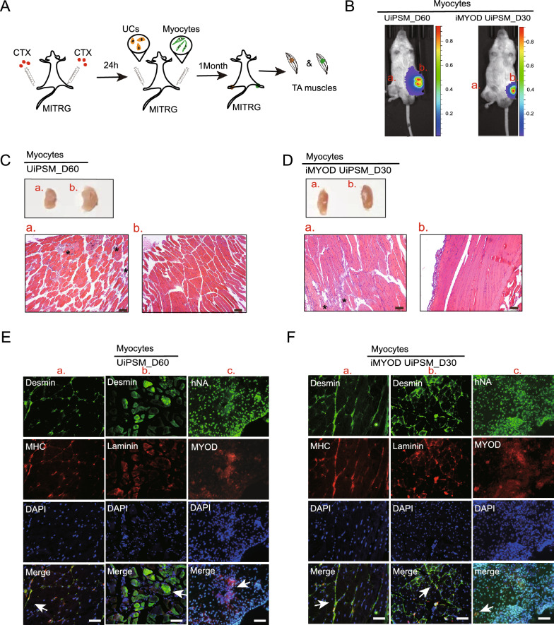
Transplantation of UiPSM and iMYOD UiPSM cells derived human myocytes in muscle injury model. A Schematic overview of the transplantation methodology for UiPSM and iMYOD UiPSM cell-derived human skeletal myocytes into the TA muscle of MITRG mice, following treatment with cardiotoxin (CTX) for 24 h. Urine cells serve as a negative control and are transplanted into the left tibialis anterior muscle. B Bioluminescence imaging (BLI) signal captured at the right tibialis anterior graft site in a representative MITRG mouse treated with CTX, 1 month after transplantation. UCs transplanted into the left TA muscle in (a), as a negative control. UiPSM derived myocytes transplanted into the rright TA muscle in (b). C Morphological characteristics of TA muscle tissue in after transplanted UiPSM-derive myocytes and UCs. H&E staining of longitudinal sections of TA muscles showed the aggregation of inflammatory factors (asterisk) could still be seen locally in the left tibial anterior muscle after transplanting UCs in (a). H&E staining of longitudinal sections of TA muscles after transplanted UiPSM differentiated into myocytes at day 60 in (b). Scale bars, 100 µm. D Morphological characteristics of TA muscle tissue in after transplanted iMYOD UiPSM-derive myocytes and UCs. H&E staining of longitudinal sections of TA muscles showed the local aggregation of inflammatory factors (asterisk)in the left tibial anterior muscle after transplanting UCs in (a). H&E staining of longitudinal sections of TA muscles after transplanted iMYOD UiPSM-derived myocytes at day 30 in (b). Scale bars, 100 µm. E TA muscle from UiPSM-derived myocytes evaluated for the expression of myocyte-specific markers. Longitudinal section showed the colocalization of Desmin (DES) and Myosin Heavy Chain (MHC) (arrows in (a.)). Transversal section showed the colocalization of Desmin and Laminin (arrows in (b.)). Transversal section showed the colocalization of human nuclei antibody (hNA) and MYOD (arrows in (c.)). Scale bars, 100 µm. F TA muscle from iMYOD UiPSM-derived myocytes evaluated for the expression of myocyte-specific markers. Colocalization of Desmin (DES) and Myosin Heavy Chain (MHC) (arrows in (a.), and colocalization of Desmin and Laminin (arrows in (b.)) were shown in Longitudinal sections. Transversal section showed the colocalization of human nuclei antibody (hNA) and MYOD (arrows in (c.)). Scale bars, 100 µm

