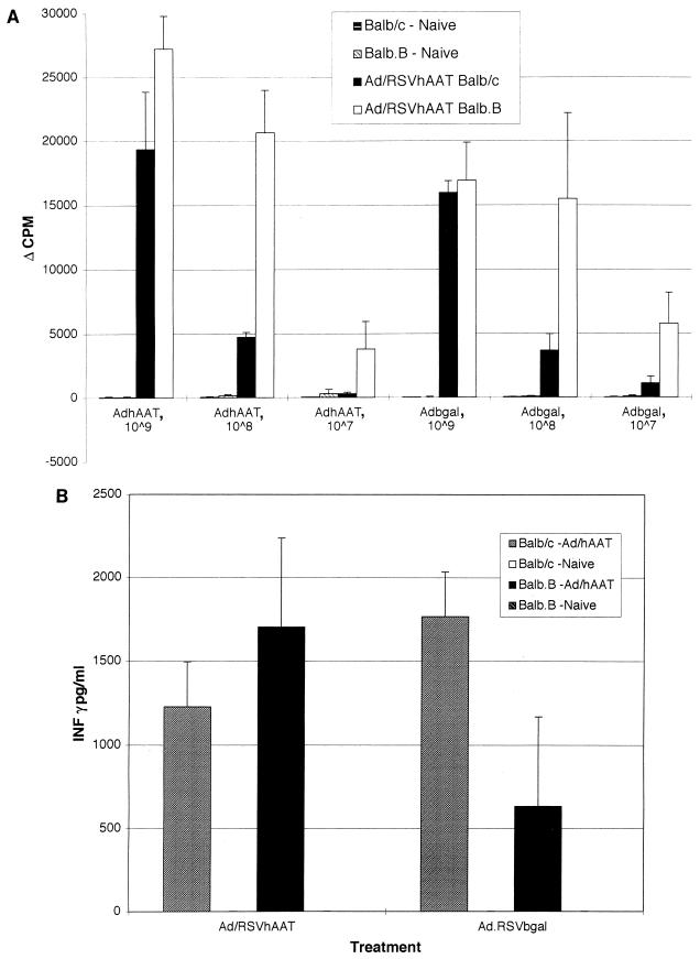
FIG. 7.
Splenocyte proliferation and IFN-γ production following infusion of Ad/RSVhAAT in Balb/c and Balb.B mice. Splenocytes were obtained from mice 15 days after the administration of 5 × 10⁹ PFU of Ad/RSVhAAT or from mice not previously exposed to adenovirus. The cells were cultured in replicates of four with medium alone, anti-murine CD3 antibody, or the indicated amounts of UV-inactivated Ad/RSVhAAT (AdhAAT), Ad/RSVβgal (Adbgal), serum containing hAAT (calibrator serum 4), or serum from an hAAT-deficient patient (null) for 72 h. Following this incubation, [3H]thymidine was added and the cells were incubated for an additional 24 h. After this final incubation, the cells were harvested, and [3H]thymidine uptake was determined and plotted as Δcpm between unstimulated and stimulated samples (A). The error bars represent standard deviations. Proliferation results for the different groups of splenocytes for unstimulated and anti-CD3-stimulated cells were similar in all groups and are not shown. No proliferation was seen following incubation with calibrator or null patient serum (not shown). Secretion of IFN-γ (B) was determined for the same splenocytes cultured in replicates of four with medium alone, anti-murine CD3 antibody, or the indicated amounts of UV-inactivated Ad/RSVhAAT or Ad/RSVβgal for 72 h. After this incubation, cell supernatants were pooled from each replicate, and the amount of IFN-γ was determined in triplicate by ELISA. The error bars represent standard errors of the means. No IFN-γ secretion was detected in the naive mice or mice treated with calibrator or null patient serum (not shown). The results for naive and unstimulated cells with the anti-CD3 antibody were similar and are not shown.

