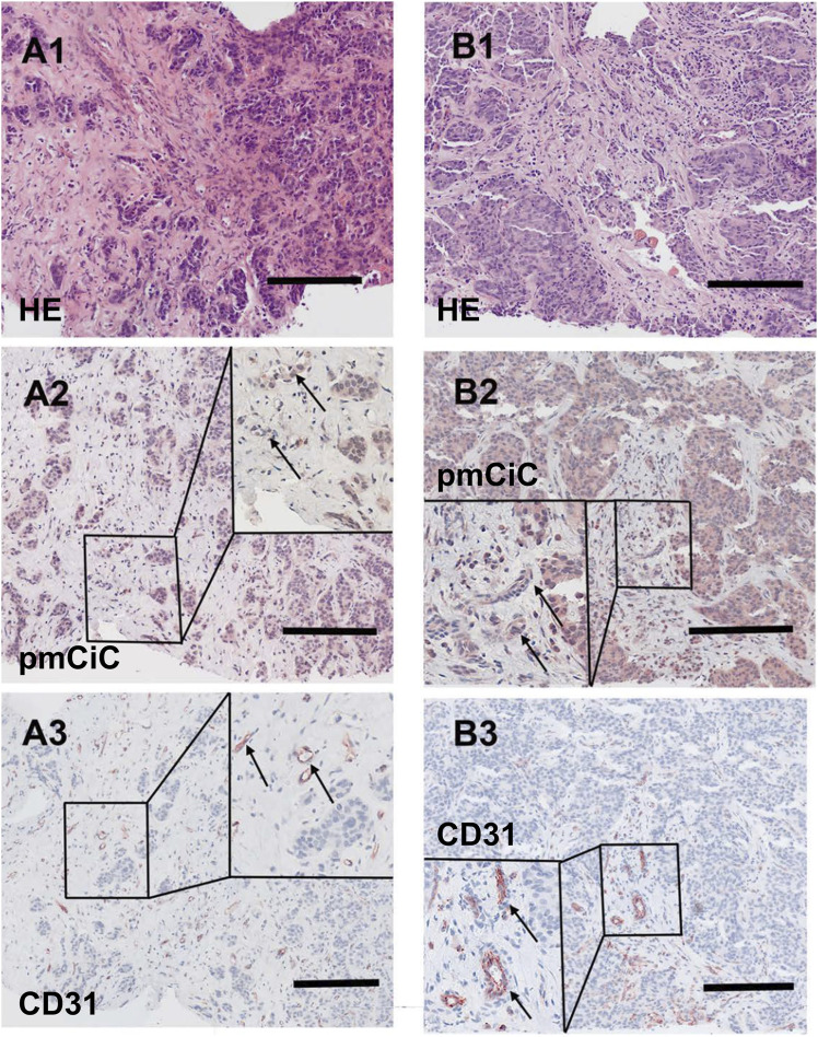
FIGURE 6.
Histological staining of a liver metastasis of a ductal breast cancer and a liver metastasis of a prostate cancer. [(A1–A3); scale bar 100 µm] Liver metastasis of a ductal breast cancer [(A1); HE] shows little nest of atypical infiltrating cancer cells with a desmoplastic stroma expressing pmCiC in tumour cells (A2) as well as some associated small vessels (inset; →; magnification 3 - fold). In (A3) serial section of the metastasis shows tumour associated vessels stained for CD31 (inset; →; magnification 3 - fold). [(B1–B3); scale bar 100 µm] Liver metastasis of a prostate cancer [(B1); HE] displaying a pseudoglandulär atypical cancer with a desmoplastic stroma expressing pmCiC in tumour cells (B2) and few tumour-associated vessels (inset; →; magnification 3 - fold). In (B3) a serial section of the specimen was stained with CD31 to highlight the tumour associated vessels (inset; →; magnification 3 - fold). Images provide an overview and details at higher magnification (inserts; magnification 3 - fold); corresponding size bars are included. Tissues shown in (A2, A3, B2, B3) were counterstained with hematoxylin.

