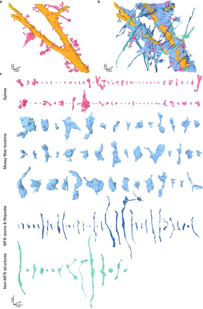
Extended Data Fig. 5. Structural characterization of the local input field in a CA3 pyramidal neuron proximal dendrite.
a, 3D-rendering of the CA3 pyramidal neuron proximal dendrite in Fig. 3g based on coCATS data. The dendritic shaft is colored in gold, spines (nspines = 68) are labeled in magenta. b, 3D-rendering of the same dendrite as in a, with associated cellular structures color-coded by identity, as inferred from morphology: MFBs (nMFBs = 43, light blue), axons and filopodia of MFBs (naxons/filopodia = 38, dark blue), structures in synaptic contact with the main dendrite, not identifiable as MFB-related structures (nnon-MFB = 14, turquoise). c, 3D-renderings of all structures reconstructed in b. Reconstruction was performed on n = 1 dataset.

