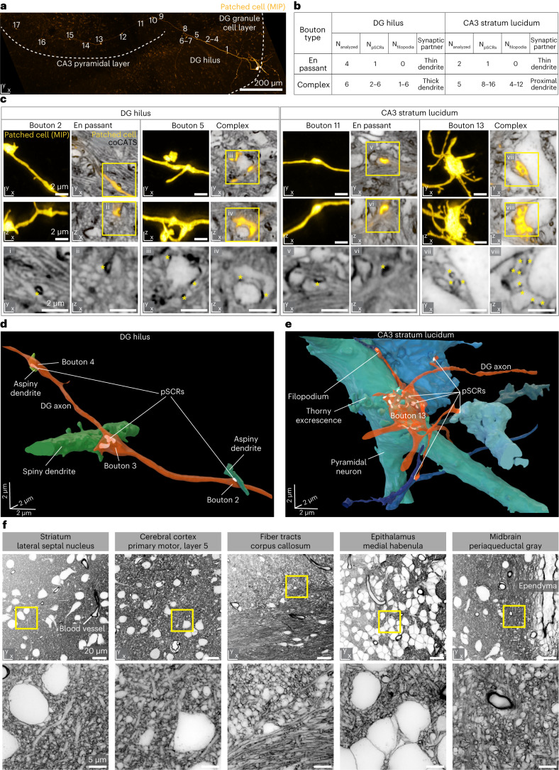
Fig. 4. Tissue architecture and single-cell output structure at individual synapse level across brain regions.
a, MIP of a whole-cell patch-clamped and biocytin-filled DG granule cell in organotypic hippocampal slice (confocal, visualized with AF594-streptavidin). Seventeen conspicuous boutons are marked along the main axon’s trajectory, projecting as mossy fiber from the DG granule cell layer through the hilus to the CA3 stratum lucidum. b, Characteristics of analyzed synaptic boutons. c, Single xy and xz planes of four example super-resolved volumes comprising specific synapses as marked in a, with coCATS (gray, z-STED, STAR RED-NHS, N2V) revealing local microenvironment of the positively labeled mossy fiber (yellow, z-STED, N2V) (raw data: Supplementary Fig. 4). Bottom: magnified views of the coCATS channel with asterisks indicating pSCRs used to identify synaptic partners. pSCRs were segmented with the same model as in Fig. 2j,k, followed by manual proofreading. d,e, 3D renderings of two axon stretches with boutons, pSCRs and synaptically connected structures in DG hilus and CA3 stratum lucidum. coCATS labeling in combination with functional recordings is representative of experiments in n = 6 organotypic slices. Following the axon trajectory with 3D reconstruction was done for n = 1 sample, with bouton characteristics extracted from a total of Nanalyzed = 17 boutons imaged across multiple volumes along the axon. f, Architecture of various regions in near-natively preserved brain revealed by coCATS with in vivo microinjection. Organization of cell bodies, dendrites, axons, synapses, ependyma around liquor spaces and blood vessels is visible. Top: confocal; bottom: xy-STED. Images represent raw data from n = 5 brain slices obtained from n = 2 independent biological specimens with in vivo microinjection into LV and primary motor cortex, respectively. They are representative of coCATS in vivo microinjection in n = 10 and n = 4 animals for LV and cortical microinjection, respectively.

