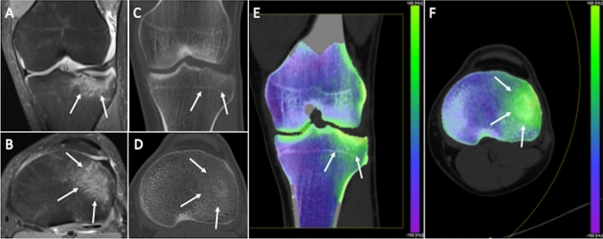
Fig. 3.
Traumatic tibial fracture in a 32-year-old woman. Coronal (a) and axial (b) fat-suppressed proton density weighted images show diffuse bone edema of lateral tibial plateau (arrows). Coronal reformat (c) and axial (d) standard CT images seem to show bone impaction of lateral tibial plateau as subtle subchondral hyperdensity (arrows), while the corresponding coronal (e) and axial (f) virtual noncalcium DECT images clearly demonstrate bone edema (arrows)

