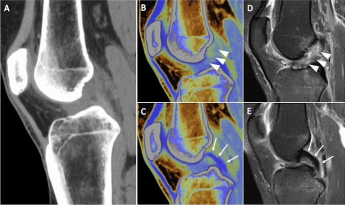
Fig. 4.
Anterior cruciate ligament tear in a 42-year-old man after knee sprain. Sagittal reformat of standard knee CT (a) is not helpful for describing clearly cruciate ligaments status. Sagittal virtual noncalcium DECT images allow the identification of anterior cruciate ligament injury (b, arrowheads), with normal appearance of posterior cruciate ligament (c, arrows), as proven by the corresponding sagittal fat-suppressed proton density weighted MRI images (d and e, respectively)

