Figure 1.
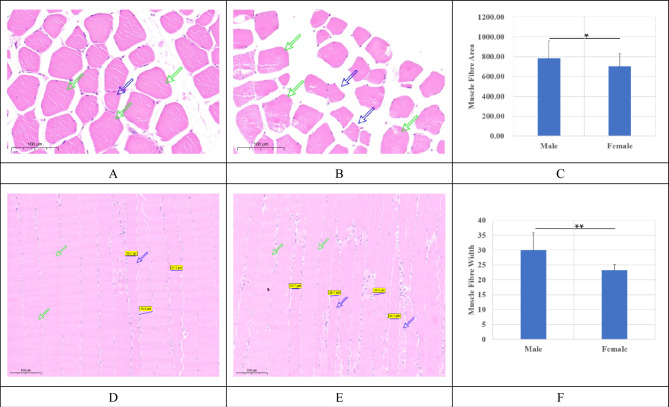
Histological staining for leg muscles from male and female geese. (A) and (D) were from male geese while (B) and (E) were for female geese. A/B and D/E were transections and longisections, respectively. The black horizontal lines in A/B/D/E were the 50 μm plotting scales. (C) were the areas of muscle fibers from the cross sections (μm2) between male and female geese. (D) were the width for the leg muscle fibers from longisections (μm) between male and female geese.

