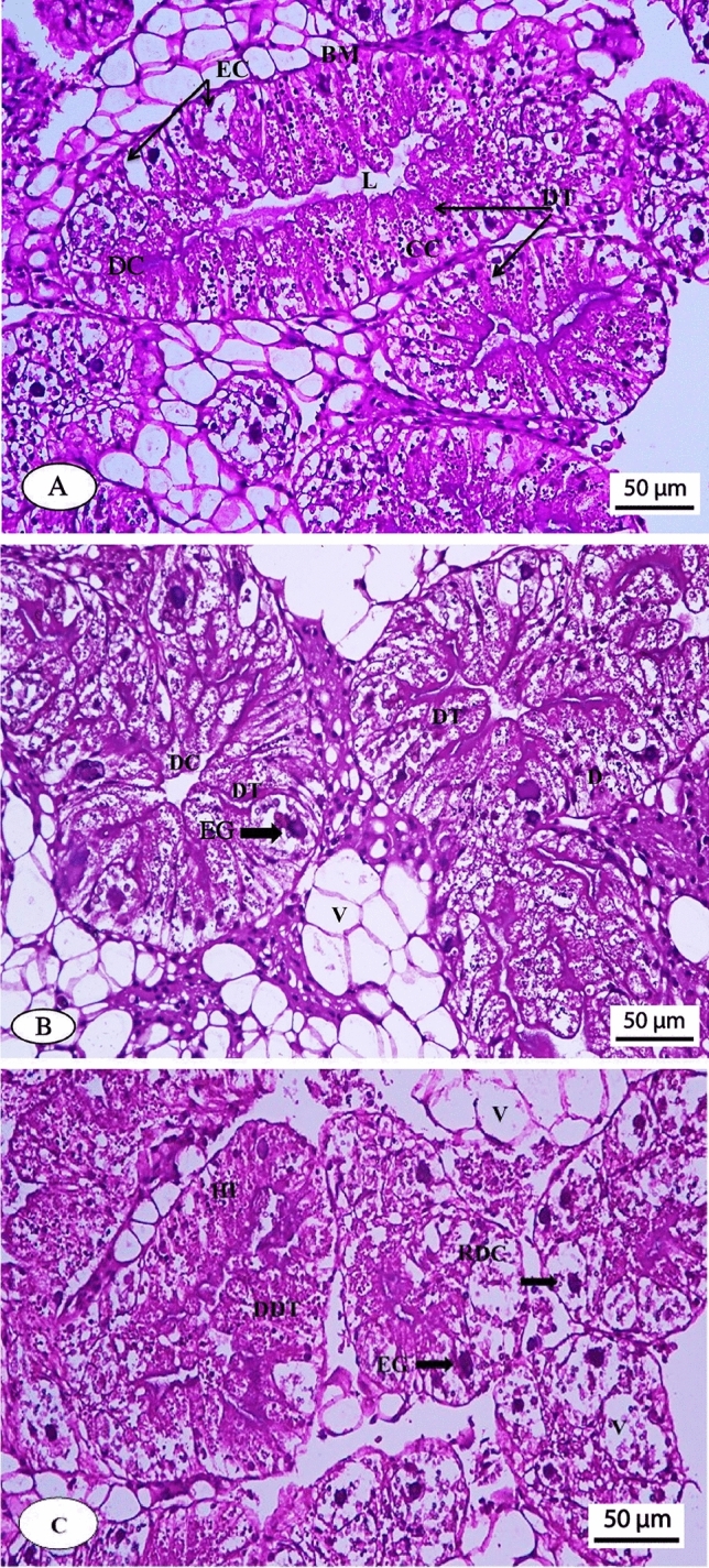
Figure 4.

Photomicrograph of the normal hepatopancreas of Theba pisana snail (A), hepatopancreas of the snail treated with 0.3 µg/ml azoxystrobin (B) and hepatopancreas of the snail treated with 3 µg/ml azoxystrobin (C). Scale bar (A, B and C): 50 μm. DC, digestive cells; DT, digestive tubules; EC, excretory cells; L, lumen; C, calcium cells; BM, bilayer membrane; V, vacuoles; HI, hemocyte infiltration; EG, excretory granules; DDT, destructed digestive tubules; RDC, ruptured digestive cells.
