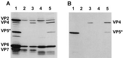
FIG. 1.
SDS-PAGE and Western blot analysis of VLP preparations. The proteins in purified SA11 (lane 1) or VLPs (G3 2/6/7-VLPs [lane 2], G3 2/4/6/7-VLPs [lane 3]), G1 2/6/7-VLPs [lane 4], and G1 2/4/6/7-VLPs [lane 5]), made in infected insect cells and purified, were separated by SDS-PAGE, transferred to nitrocellulose, and detected with a hyperimmune anti-SA11 mouse serum (A) or an anti-VP4 monoclonal antibody, 5E4 (B). Locations of the individual proteins are shown at the left and right.

